motion anatomy
Motion Is holistic.
Chapter 3
Pick up a glass of water.
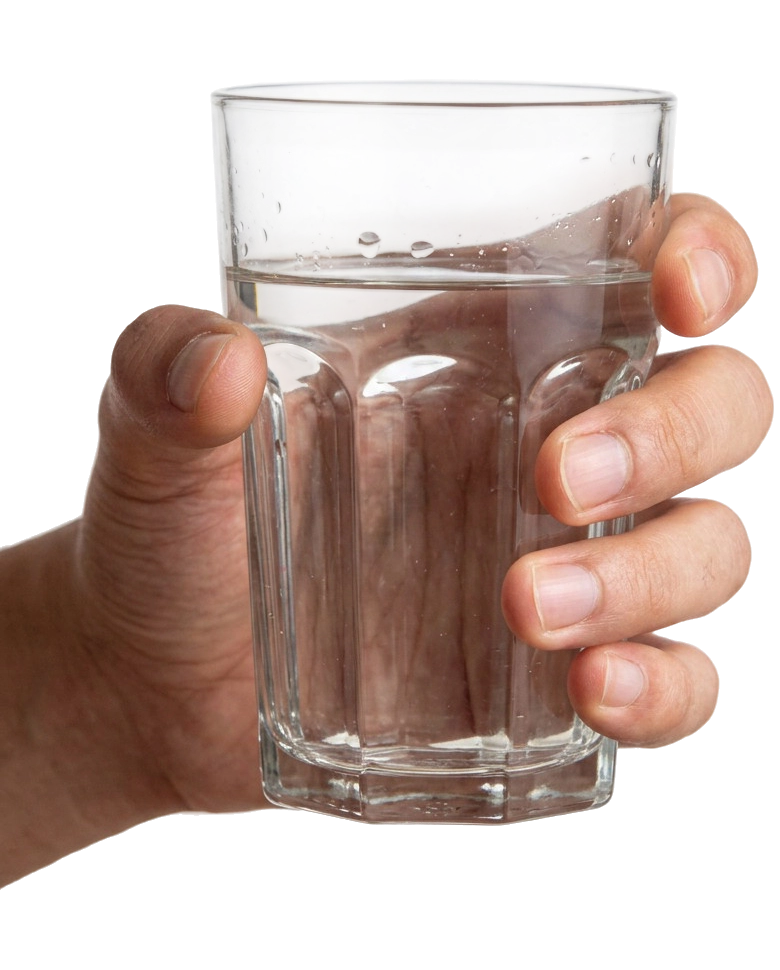
Before your hand reaches the glass, your eyes locate it. Your nervous system plans the reach. The muscles of your shoulder, arm, and hand fire in the precise sequence needed to extend, grasp, and lift (Castiello, 2005). Your respiratory rate adjusts to support the effort (Dempsey et al., 2020). Your heart maintains the cardiac output needed to deliver oxygen and substrate to the working tissues. Your circulatory system dilates the vessels supplying the working muscles and constricts those supplying tissues that can wait (Joyner & Casey, 2015). Your endocrine system helps maintain a baseline state: insulin to regulate glucose, the diurnal rhythm of cortisol, the pulsatile release of growth hormone for tissue maintenance, and the release of aldosterone for fluid and electrolyte balance all play a role (Hall & Hall, 2020). Your skin's mechano- and thermoreceptors register the glass's surface, weight, and temperature. They feed that information back to your nervous system in real time (Johansson & Flanagan, 2009). Your fascial network transmits force from your fingers through your wrist, forearm, shoulder, and trunk, distributing the load (Stecco, 2015). Your skeleton positions itself to support the lift. Your lymphatic system, propelled by the muscle contractions of your arm, moves fluid that has been stagnant since your last movement (Scallan et al., 2016). Your digestive, urinary, and reproductive systems continue their work—and contribute their hormonal, metabolic, and circulatory states to whatever happens next.
You picked up a glass of water. Every organ system played a role.
This is not an exaggeration. This is reality.
What Is Motion
Before going further, a definition. The word "motion" can mean many things, and the broader its use, the harder it is to talk about with precision.
On this website, motion is an umbrella term. It encompasses any change in the position of the body or its parts. Motion ranges from cellular contraction at one end of the scale to whole-body activity at the other. It ranges from the shortening of a single sarcomere (Huxley & Niedergerke, 1954) and the propulsion of a single lymphocyte through tissue (Friedl & Weigelin, 2008) to walking, running, dancing, lifting, and throwing. It includes daily activities such as driving and work, as well as therapeutic treatment and exercise, resistance training, cardiovascular exercise, and sports. And it includes involuntary motion: breathing, digestion, vascular pulsation, and lymphatic propulsion.
Defining motion broadly is not a stylistic choice. It is a clinical necessity. The cellular processes that produce a single muscle contraction are the same ones that, when summed and coordinated, produce a marathon. There is no clean line between "small motion" and "large motion." It is one phenomenon, occurring at many scales simultaneously. Any clinical reasoning that treats them as separate fails to account for how the body actually works.

The Organ Systems And Their Motion-Related Functions
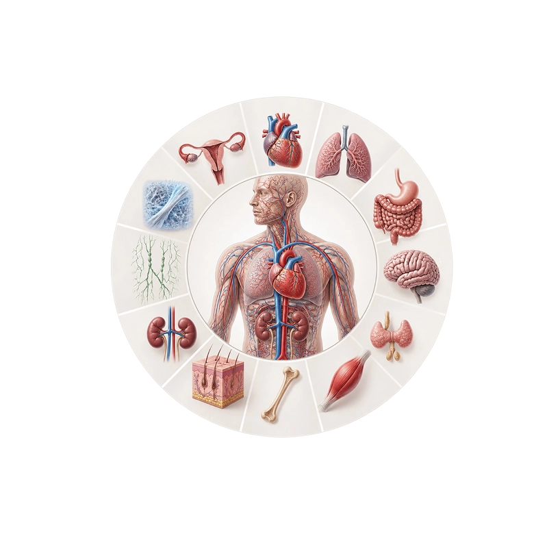
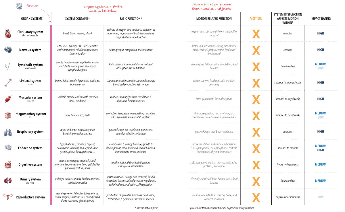
The “motion is holistic” table below shows the role of each organ system during motion. It is useful to keep close. What follows is a tour through it with the supporting literature attached.
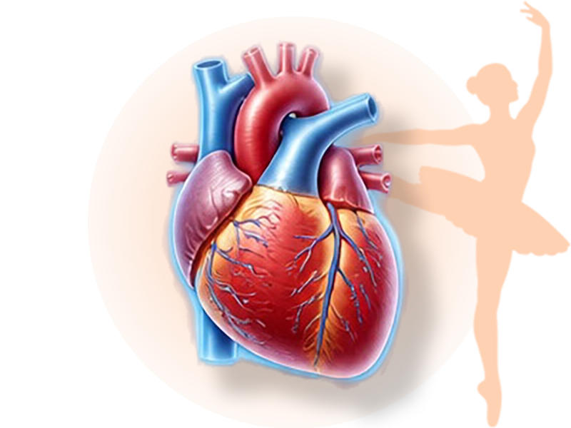
The circulatory system delivers the oxygen and substrates that working tissues require, removes metabolic byproducts, and regulates body temperature during sustained activity (Joyner & Casey, 2015; Rowell, 1993). The heart drives the system; cardiac dysfunction has motion consequences well beyond the cardiovascular complaint itself (Duncker & Bache, 2008). Motion returns the favor: muscular contraction, particularly in the calf, augments venous return — the soleus is sometimes called a "second heart" for this reason (Rowell, 1993; Recek, 2013). Dysfunction shows up in motion within minutes. Impact rating: high.
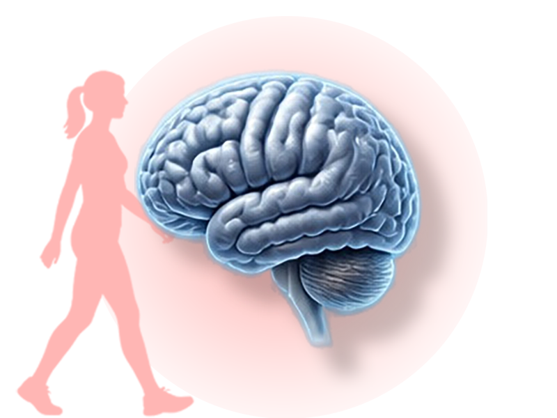
The nervous system provides the motor planning, motor unit recruitment, firing rate control, proprioceptive feedback, and motor control that turn intention into coordinated movement (Proske & Gandevia, 2012; Tuthill & Azim, 2018). Dysfunction here changes motion in seconds. Impact rating: high. Without intact neural function, no motion happens — or what happens is uncoordinated, mistimed, or absent.

The fascial system transmits force throughout the body, provides the medium through which the lymphatic system drains, and actively participates in mechanical and neurological signaling (Myers, 2020; Stecco, 2015; Schleip, 2003; Ingber, 1997; Wiig & Swartz, 2012; Benias et al., 2018). Dysfunction affects motion on a timescale of minutes to months. Impact rating: difficult to assign categorically, but the evidence increasingly supports a substantial role.
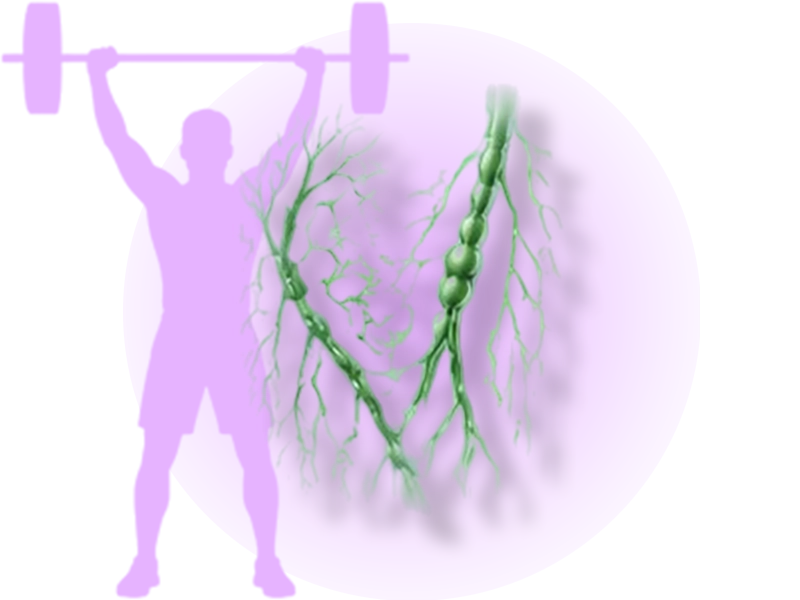
The lymphatic system handles tissue repair, regulates inflammation, and balances fluid during and after movement (Hespe et al., 2016). It depends on motion for its flow — there is no central pump, so lymph moves by muscular contraction, breathing, and tissue compression (Scallan et al., 2016). Dysfunction shows up on a timescale of hours to days. Impact rating: medium-low for acute motion, but the cumulative effect of impaired lymphatic function on tissue health and pain is substantial.
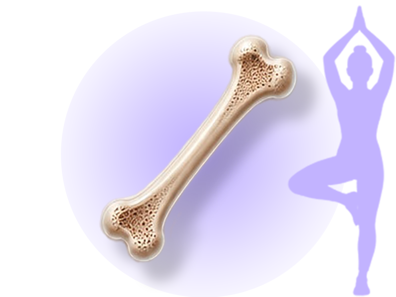
The skeletal system provides the levers, load transmission, and joint geometry that enable organized motion (Neumann, 2017). Dysfunction here affects motion on a timescale of seconds (an acute joint sprain changes motion immediately) to months or years (a chronic degenerative joint condition reshapes movement over the long term). The impact rating is high. No system more directly defines the architecture of how a body moves.
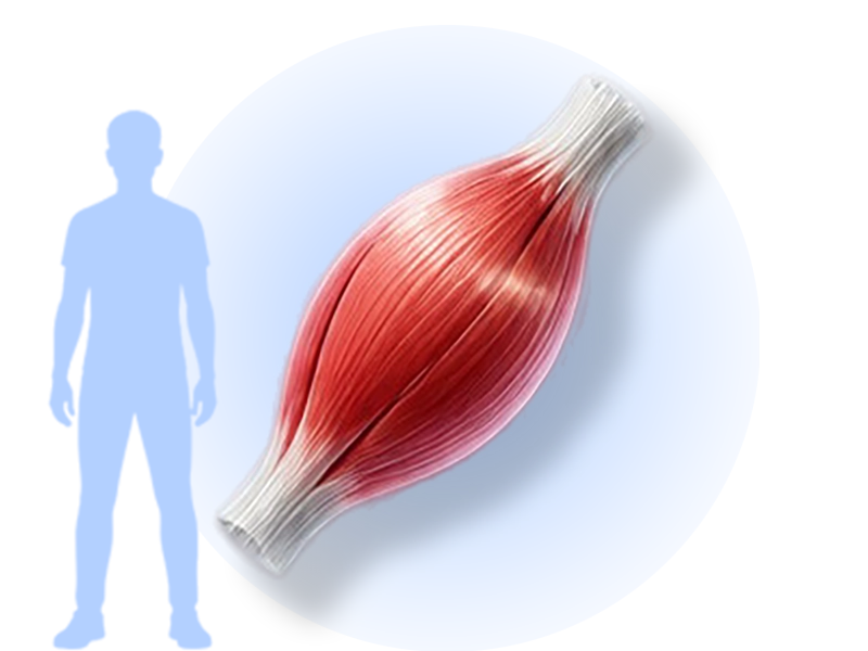
The muscular system generates and absorbs the forces that produce motion (Fitts, 1994; Gordon et al., 1966; Huxley & Niedergerke, 1954). It is also a metabolically active organ in its own right, secreting myokines that affect bone, fat, brain, and immune function (Pedersen & Febbraio, 2012). Dysfunction affects motion on a timescale of seconds to weeks. Impact rating: high.
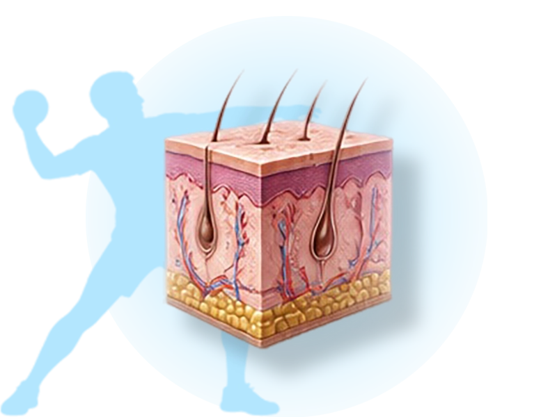
The integumentary system does more during motion than you might think. It is the body's thermoregulatory interface, dissipating heat through sweating during sustained activity (Romanovsky, 2014; Baker, 2019). It provides sensory input through its dense mechanoreceptor network, telling the nervous system what your body is in contact with and how (Johansson & Flanagan, 2009). It protects underlying structures from mechanical insult during movement. Its dysfunction shows up in motion on a timescale of minutes to weeks — a sunburn changes how you move within hours; a chronic skin condition shifts movement patterns over weeks. The impact rating is medium-low, reflecting that the skin's role is supportive rather than primary, though it is real.
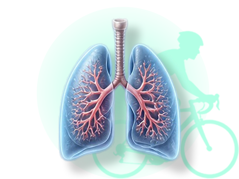
The respiratory system handles gas exchange and acid-base regulation during motion (Dempsey et al., 2020; Romer & Polkey, 2008). Dysfunction affects motion within minutes. Impact rating: high. The respiratory muscles themselves generate movement, and breathing patterns affect autonomic state, fascial mobility through the diaphragm's central role in trunk function (Bordoni & Zanier, 2013), and vagal tone (Porges, 2007). Motion changes breathing, and breathing changes motion.
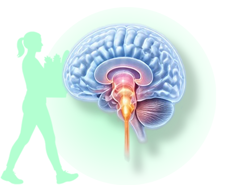
The endocrine system modulates motion through acute hormonal responses (epinephrine, norepinephrine, cortisol) and chronic adaptations (testosterone, thyroid hormones, growth factors) (Kraemer & Ratamess, 2005; Hackney & Lane, 2015; Athanasiou et al., 2023). Dysfunction affects motion on a timescale of seconds (acute stress response) to months (chronic endocrine dysregulation). Impact rating: medium-high.
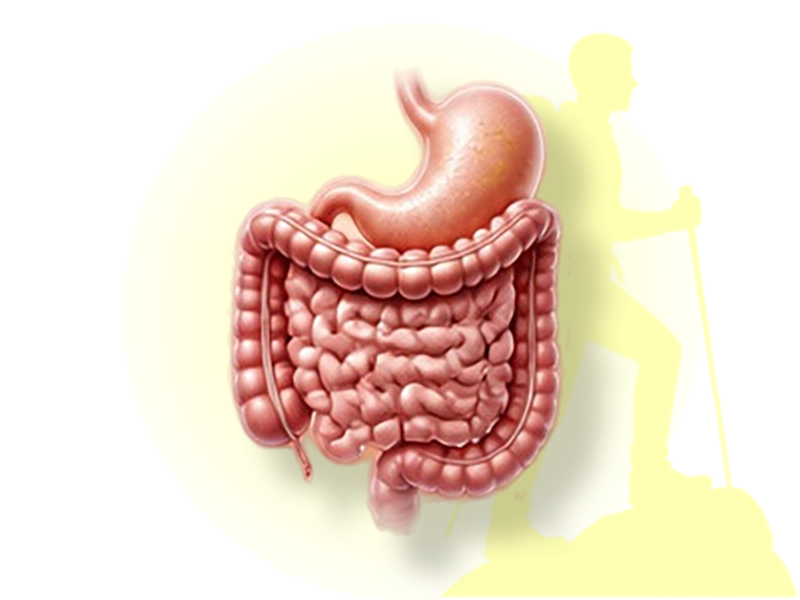
The digestive system provides the substrate (glucose, fatty acids, proteins) and the hydration that sustained motion requires (Ormsbee et al., 2014; Jeukendrup, 2017). Dysfunction shows up in motion over hours to weeks. Impact rating: medium.

The renal system maintains electrolyte and acid-base balance, fluid balance, and waste clearance on which motion depends (Poortmans & Ouchinsky, 2006). Dysfunction affects motion on a timescale of hours to days. Impact rating: medium.

The reproductive system contributes through the effects of sex hormones on muscle, bone, and connective tissue (Chidi-Ogbolu & Baar, 2019; Hunter et al., 2023; Handelsman et al., 2018). Dysfunction affects motion on a timescale of days to months. Impact rating: low for acute motion, but the cumulative effects on tissue quality and injury susceptibility are documented.
Why This Matters Clinically
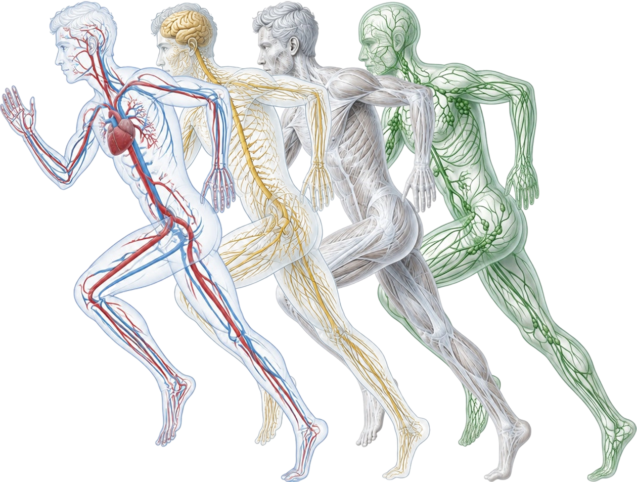
The list above is exhaustive in one sense: every organ system participates in every motion. It is finite in another: there are eleven systems plus fascia, and that’s it, and that’s all. The twelve-system picture is not a claim of vague holism. It is a specific, all-encompassing description of what works whenever a body moves.
For a practitioner, this has three immediate consequences.
The first is that any intervention you apply to a body affects more than the structures you intend to treat or train (Sueki et al., 2013). When you mobilize or move a joint, you change skeletal mechanics, the proprioceptive input the joint sends to the nervous system, the lymphatic flow from surrounding tissue, the circulatory pattern in the region, and the person's autonomic state. Although you hone in on certain parts of certain organ systems, you can’t avoid changing all of them. They are intertwined.
The second is that impaired function in any system shows up in motion, though not always visibly. A client with dysfunctional digestion moves differently from one with a healthy system (Lima et al., 2025). Poorly managed chronic physical, mental, and emotional stress also changes a client's movement compared to well-managed, intermittent stress (Metz, 2007). Some changes are obvious and easy to spot. Many are subtle shifts in coordination, timing, or autonomic state and may persist for months or years before becoming visible. By the time the change is unmistakable, the body has often adapted, turning a small problem into a widespread, difficult-to-resolve one. The absence of visible change in motion does not mean there is no problem.
The third — and the one this website is built around — is that motion itself is therapeutic across all systems. When a client moves well, every system benefits. When motion is restored to a system that has been moving poorly, the positive effects ripple beyond the immediate complaint. This is why motion anatomy organizes gross anatomy by motion: because motion is the common denominator across every organ system, and reorganizing anatomy around it is the most direct route to clinical leverage.
Where This Leaves Us
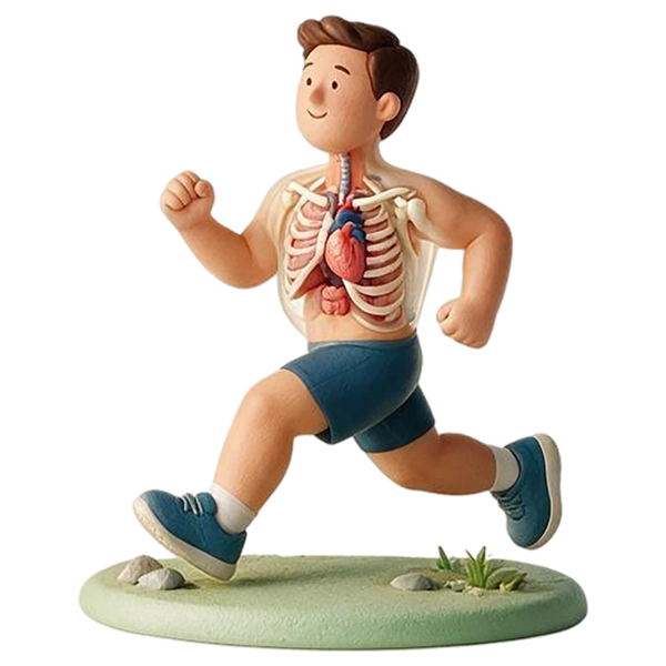
Motion is holistic. Eleven organ systems plus fascia participate in every motion you produce, large or small, voluntary or involuntary. The literature supporting this is in the standard references and the peer-reviewed journals.
The next chapter introduces the anatomical planes of motion, and the following chapter explains that motion anatomy is gross anatomy combined with motion. It’s the foundation for the clinical framework on which the rest of this website is built.
Next Chapter
Summary & References
Motion is holistic
- A single act — picking up a glass — simultaneously engages vision, nervous planning, muscle, respiration, circulation, endocrine state, skin receptors, fascia, skeleton, lymphatics, and the visceral systems.
- The point made concrete: no motion, however small, is the work of one system.
Definition of motion
- Motion is defined broadly: any change in position of the body or its parts, from a single sarcomere shortening or a lymphocyte propelled through tissue to walking, lifting, and sport, including involuntary motion like breathing and digestion.
- The broad definition is a clinical necessity, not a style choice: the same cellular processes that make one contraction make a marathon, so there's no clean line between "small" and "large" motion.
Motion and organ systems
- A system-by-system tour of each organ system's motion role, with the supporting literature attached and an impact rating for each.
- Examples: circulation delivers oxygen and clears byproducts (and motion repays it — the calf as a "second heart"); the nervous system turns intention into coordinated movement, and its dysfunction changes motion in seconds.
- The reciprocity is explicit: systems enable motion, and motion in turn serves the systems.
Why this matters clinically
- The eleven-systems-plus-fascia picture is finite and specific, not vague holism — it names exactly what is at work whenever a body moves.
- Three consequences: any intervention affects more than the structure under your hand; impaired function in any system shows up in motion (often subtly, sometimes for months before it's visible); the body doesn't separate the effects you intended from the ones you didn't.
Reference list chapter 3
- Athanasiou, N., Bogdanis, G. C., & Mastorakos, G. (2023). Endocrine responses of the stress system to different types of exercise. Reviews in Endocrine and Metabolic Disorders, 24(2), 251–266. https://doi.org/10.1007/s11154-022-09758-1
- Baker, L. B. (2019). Physiology of sweat gland function: The roles of sweating and sweat composition in human health. Temperature, 6(3), 211–259. https://doi.org/10.1080/23328940.2019.1632145
- Benias, P. C., Wells, R. G., Sackey-Aboagye, B., Klavan, H., Reidy, J., Buonocore, D., Miranda, M., Kornacki, S., Wayne, M., Carr-Locke, D. L., & Theise, N. D. (2018). Structure and distribution of an unrecognized interstitium in human tissues. Scientific Reports, 8, 4947. https://doi.org/10.1038/s41598-018-23062-6
- Bordoni, B., & Zanier, E. (2013). Anatomic connections of the diaphragm: Influence of respiration on the body system. Journal of Multidisciplinary Healthcare, 6, 281–291. https://doi.org/10.2147/JMDH.S45443
- Castiello, U. (2005). The neuroscience of grasping. Nature Reviews Neuroscience, 6(9), 726–736. https://doi.org/10.1038/nrn1744
- Chidi-Ogbolu, N., & Baar, K. (2019). Effect of estrogen on musculoskeletal performance and injury risk. Frontiers in Physiology, 9, 1834. https://doi.org/10.3389/fphys.2018.01834
- Dempsey, J. A., La Gerche, A., & Hull, J. H. (2020). Is the healthy respiratory system built just right, overbuilt, or underbuilt to meet the demands imposed by exercise? Journal of Applied Physiology, 129(6), 1235–1256. https://doi.org/10.1152/japplphysiol.00444.2020
- Duncker, D. J., & Bache, R. J. (2008). Regulation of coronary blood flow during exercise. Physiological Reviews, 88(3), 1009–1086. https://doi.org/10.1152/physrev.00045.2006
- Fitts, R. H. (1994). Cellular mechanisms of muscle fatigue. Physiological Reviews, 74(1), 49–94. https://doi.org/10.1152/physrev.1994.74.1.49
- Friedl, P., & Weigelin, B. (2008). Interstitial leukocyte migration and immune function. Nature Immunology, 9(9), 960–969. https://doi.org/10.1038/ni.f.212
- Gordon, A. M., Huxley, A. F., & Julian, F. J. (1966). The variation in isometric tension with sarcomere length in vertebrate muscle fibres. The Journal of Physiology, 184(1), 170–192. https://doi.org/10.1113/jphysiol.1966.sp007909
- Hackney, A. C., & Lane, A. R. (2015). Exercise and the regulation of endocrine hormones. Progress in Molecular Biology and Translational Science, 135, 293–311. https://doi.org/10.1016/bs.pmbts.2015.07.001
- Hall, J. E., & Hall, M. E. (2020). Guyton and Hall textbook of medical physiology (14th ed.). Elsevier.
- Handelsman, D. J., Hirschberg, A. L., & Bermon, S. (2018). Circulating testosterone as the hormonal basis of sex differences in athletic performance. Endocrine Reviews, 39(5), 803–829. https://doi.org/10.1210/er.2018-00020
- Hespe, G. E., Kataru, R. P., Savetsky, I. L., García Nores, G. D., Torrisi, J. S., Nitti, M. D., Gardenier, J. C., Zhou, J., Yu, J. Z., Jones, L. W., & Mehrara, B. J. (2016). Exercise training improves obesity-related lymphatic dysfunction. The Journal of Physiology, 594(15), 4267–4282. https://doi.org/10.1113/JP271757
- Hunter, S. K., Angadi, S. S., Bhargava, A., Harper, J., Hirschberg, A. L., Levine, B. D., Moreau, K. L., Nokoff, N. J., Stachenfeld, N. S., & Bermon, S. (2023). The biological basis of sex differences in athletic performance: Consensus statement for the American College of Sports Medicine. Medicine & Science in Sports & Exercise, 55(12), 2328–2360. https://doi.org/10.1249/MSS.0000000000003300
- Huxley, A. F., & Niedergerke, R. (1954). Structural changes in muscle during contraction: Interference microscopy of living muscle fibres. Nature, 173(4412), 971–973. https://doi.org/10.1038/173971a0
- Ingber, D. E. (1997). Tensegrity: The architectural basis of cellular mechanotransduction. Annual Review of Physiology, 59, 575–599. https://doi.org/10.1146/annurev.physiol.59.1.575
- Jeukendrup, A. E. (2017). Periodized nutrition for athletes. Sports Medicine, 47(Suppl. 1), 51–63. https://doi.org/10.1007/s40279-017-0694-2
- Johansson, R. S., & Flanagan, J. R. (2009). Coding and use of tactile signals from the fingertips in object manipulation tasks. Nature Reviews Neuroscience, 10(5), 345–359. https://doi.org/10.1038/nrn2621
- Joyner, M. J., & Casey, D. P. (2015). Regulation of increased blood flow (hyperemia) to muscles during exercise: A hierarchy of competing physiological needs. Physiological Reviews, 95(2), 549–601. https://doi.org/10.1152/physrev.00035.2013
- Kraemer, W. J., & Ratamess, N. A. (2005). Hormonal responses and adaptations to resistance exercise and training. Sports Medicine, 35(4), 339–361. https://doi.org/10.2165/00007256-200535040-00004
- Lima, F. L. L. C., Barreto, T. do N. P., Melo, T. M. de S., da Silva, M. L., Assunção, M. T. B., de Alencar, G. G., da Silva, G. A. P., & de Siqueira, G. R. (2025). Postural deviation patterns, trunk mobility restriction and spinal musculoskeletal dysfunctions in chronic gastritis. Journal of Bodywork and Movement Therapies, 42, 78–86. https://doi.org/10.1016/j.jbmt.2024.11.036
- Metz, G. A. (2007). Stress as a modulator of motor system function and pathology. Reviews in the Neurosciences, 18(3–4), 209–222. https://doi.org/10.1515/REVNEURO.2007.18.3-4.209
- Myers, T. W. (2020). Anatomy trains: Myofascial meridians for manual therapists and movement professionals (4th ed.). Elsevier.
- Neumann, D. A. (2017). Kinesiology of the musculoskeletal system: Foundations for rehabilitation (3rd ed.). Mosby/Elsevier.
- Ormsbee, M. J., Bach, C. W., & Baur, D. A. (2014). Pre-exercise nutrition: The role of macronutrients, modified starches and supplements on metabolism and endurance performance. Nutrients, 6(5), 1782–1808. https://doi.org/10.3390/nu6051782
- Pedersen, B. K., & Febbraio, M. A. (2012). Muscles, exercise and obesity: Skeletal muscle as a secretory organ. Nature Reviews Endocrinology, 8(8), 457–465. https://doi.org/10.1038/nrendo.2012.49
- Poortmans, J. R., & Ouchinsky, M. (2006). Glomerular filtration rate and albumin excretion after maximal exercise in aging sedentary and active men. The Journals of Gerontology Series A: Biological Sciences and Medical Sciences, 61(11), 1181–1185. https://doi.org/10.1093/gerona/61.11.1181
- Porges, S. W. (2007). The polyvagal perspective. Biological Psychology, 74(2), 116–143. https://doi.org/10.1016/j.biopsycho.2006.06.009
- Proske, U., & Gandevia, S. C. (2012). The proprioceptive senses: Their roles in signaling body shape, body position and movement, and muscle force. Physiological Reviews, 92(4), 1651–1697. https://doi.org/10.1152/physrev.00048.2011
- Recek, C. (2013). Calf pump activity influencing venous hemodynamics in the lower extremity. International Journal of Angiology, 22(1), 23–30. https://doi.org/10.1055/s-0033-1334092
- Romanovsky, A. A. (2014). Skin temperature: Its role in thermoregulation. Acta Physiologica, 210(3), 498–507. https://doi.org/10.1111/apha.12231
- Romer, L. M., & Polkey, M. I. (2008). Exercise-induced respiratory muscle fatigue: Implications for performance. Journal of Applied Physiology, 104(3), 879–888. https://doi.org/10.1152/japplphysiol.01157.2007
- Rowell, L. B. (1993). Human cardiovascular control. Oxford University Press.
- Scallan, J. P., Zawieja, S. D., Castorena-Gonzalez, J. A., & Davis, M. J. (2016). Lymphatic pumping: Mechanics, mechanisms and malfunction. The Journal of Physiology, 594(20), 5749–5768. https://doi.org/10.1113/JP272088
- Schleip, R. (2003). Fascial plasticity — A new neurobiological explanation: Part 1. Journal of Bodywork and Movement Therapies, 7(1), 11–19. https://doi.org/10.1016/S1360-8592(02)00067-0
- Stecco, C. (2015). Functional atlas of the human fascial system. Churchill Livingstone/Elsevier.
- Sueki, D. G., Cleland, J. A., & Wainner, R. S. (2013). A regional interdependence model of musculoskeletal dysfunction: Research, mechanisms, and clinical implications. Journal of Manual & Manipulative Therapy, 21(2), 90–102. https://doi.org/10.1179/2042618612Y.0000000027
- Tuthill, J. C., & Azim, E. (2018). Proprioception. Current Biology, 28(5), R194–R203. https://doi.org/10.1016/j.cub.2018.01.064
- Wiig, H., & Swartz, M. A. (2012). Interstitial fluid and lymph formation and transport: Physiological regulation and roles in inflammation and cancer. Physiological Reviews, 92(3), 1005–1060. https://doi.org/10.1152/physrev.00037.2011
Move toward a better you!
Strength
Flexibility

Biological Age
A Stronger Heart

Mental Resilience

Sharper Mind

Stronger Bones