
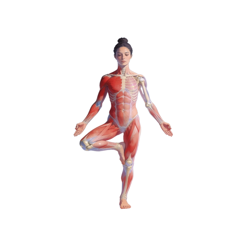
motion anatomy
Motion anatomy.

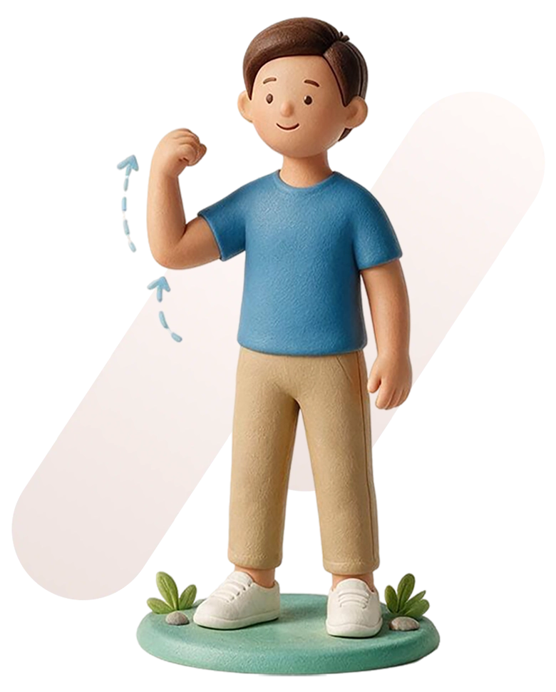
Chapter 5
Motions like extension of the sagittal knee, lateral flexion of the frontal thoracic, and rotation of the transverse shoulder involve clusters of structures from all organ systems. Motion never occurs in isolation through a single structure, region, or system. It transcends all.
Motion occurs through one or more joints simultaneously. It is generated by multiple muscles. It involves several skin areas and requires multiple peripheral nerves, spinal cord segments, arteries, veins, fascial structures, lymphatic vessels, nodes, and more (Neumann, 2017).
This is what makes combining gross anatomy and motion both challenging and clinically practical.
Organizing human anatomy by flexion-extension, abduction-adduction, and lateral-medial rotation means grouping it by all the structures that enable these motions.
That grouping is called a complex.
Gross anatomy organizes the body by region and system; motion anatomy organizes it by complex.
What Is a complex
The word "complex" means "consisting of different connecting parts," which is exactly what it is here (Oxford English Dictionary, n.d.).
In motion anatomy, a complex is a cluster of interconnected structures of the circulatory, nervous, fascial, lymphatic, muscular, skeletal, integumentary, and all other systems (see Chapter 2) grouped by a plane-specific motion they share: flexion-extension, abduction-adduction, or lateral-medial rotation (see Chapter 4).
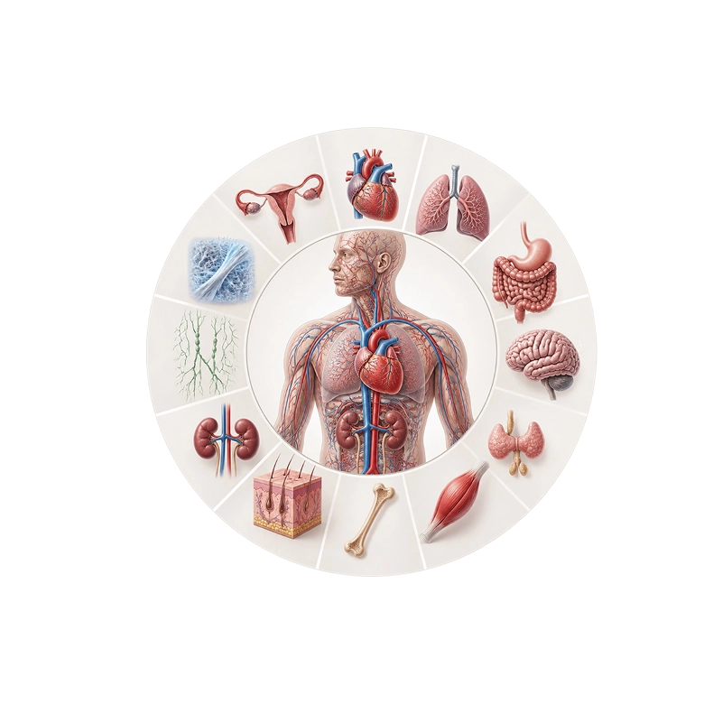
Per plane, complex, and clockwise-counterclockwise motions, arteries and their veins, peripheral nerves with related spinal segments and CNS, deep and superficial fasciae, loose connective tissues, lymphatic vessels and nodes, one or multiple joints with their capsules and ligaments, muscles, peripheral and segmentally innervated skin (Neumann, 2017), and other interconnected structures and organs of the respiratory, endocrine, digestive, urinary, and reproductive systems participate. They do so directly and indirectly (see Chapters 2 and 3).
Motion happens because everything in a complex interconnects, communicates, and does its part — motion is a team effort (see Chapters 2 and 3).
There are 19 complexes in total, ranging from the big toe to the fingers (see below).
Although neighboring complexes may share structures, each is unique. This uniqueness comes from each complex's specific combination of, for example, arteries, veins, peripheral nerves, spinal segments, fasciae, lymph vessels, joints, muscles, and related skin. No two complexes share the same combination.
complex examples
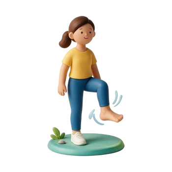
Frontal ankle
All your organ systems are active when you evert-invert your frontal ankle, but some are more involved than others. This graph shows their estimated relative involvement.
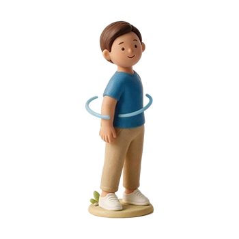
Transverse thoracic
All your organ systems are active when you left-right rotate your transverse thoracic, but some are more involved than others. This graph shows their estimated relative involvement.
complexes per plane
Each anatomical plane contains 19 complexes, most active and some passive (see below).
The sagittal plane contains 16 active and 3 passive complexes. The passive sagittal pubic symphysis, sacroiliac, and radioulnar move several degrees through movement of their neighbors.
The frontal plane has 12 active and 7 passive complexes. The passive frontal big toe, lesser toes, knee, pubic symphysis, sacroiliac, elbow, and radioulnar can move a bit through movement of their active neighbors.
Finally, the transverse plane has 11 active and 8 passive complexes. Like the other planes, the passive transverse ankle, pubic symphysis, sacroiliac, rib cage, elbow, wrist, fingers, and thumb move only a little through the movement of other complexes.

Each anatomical plane contains 19 complexes, most active and some passive.
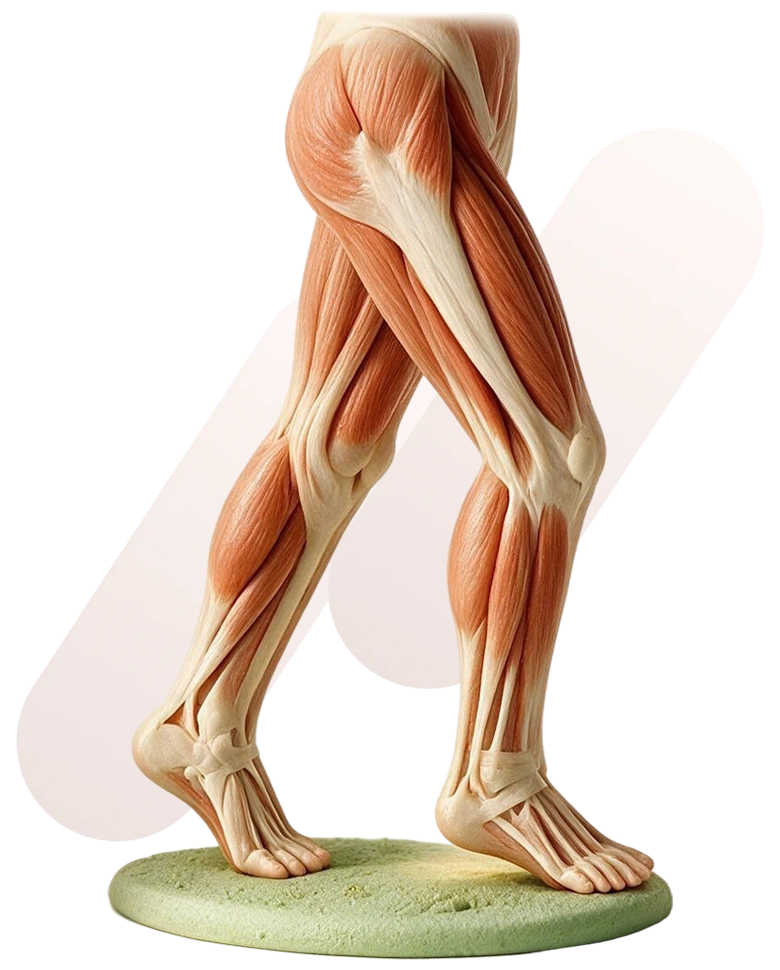
Passive and active complexes
Not every complex actively moves; some of them are passive.
A passive complex participates somewhat in its plane but lacks dedicated movers and is not designed for extensive motions in that plane.
The frontal knee is an example of a passive complex.
Although the knee moves a few degrees in the frontal plane during hip and ankle movements, increasing with flexion and load, it is not designed for true abduction-adduction. The knee lacks its own abductor-adductor muscles; its articular surfaces do not accommodate extensive abduction-adduction, and the collateral and cruciate ligaments limit and resist frontal plane movements (Neumann, 2017).
If the frontal knee does move more than it should, it can lead to an ACL or MCL injury (Neumann, 2017).
Keep in mind the opposite is also true. If a passive complex does not move at all, if the frontal knee does not allow its few degrees of abduction-adduction, pain and injury occur (Neumann, 2017). Like an active complex, a passive complex should not get stuck. Both must be functional for full range, pain-free movement.
Of all passive complexes, only the pubic symphysis, sacroiliac, and rib cage are extensively researched. Their movement in response to neighboring complexes and the details of these movements are well established (Walheim & Selvik, 1984; Sturesson et al., 2000; Beyer et al., 2014). Research on other passive complexes and their healthy motion is scarce or methodologically questionable. This is why, in motion anatomy, most passive complexes lack anatomical and clinical details and are generally left blank. I hope research will fill these gaps over time.
single- and multi-complex muscles
Because motion happens per complex, it is useful to mention how many complexes a muscle moves. In motion anatomy, we use the terms single-complex and multi-complex muscles.
Single-complex muscles belong to and move just one complex. These muscles are usually the main movers and cross one or more joints. They are often, but not always, located close to the bones in the thighs, torso, and upper arms, and more superficial in the lower legs and forearms. In contrast, multi-complex muscles involve two or more complexes. These muscles usually help control and coordinate movement. They cross two or more joints and generally run close to the surface in the thighs, torso, and upper arms, and close to the bones in the lower legs and forearms.
Examples of single-complex muscles include the vastus lateralis, gluteus medius, lumbar multifidus, and brachialis. Each one is part of and moves a specific complex: the knee, hip, lumbar, and elbow, in that order (Neumann, 2017).
The tensor fasciae latae is a multi-complex muscle because it is involved with both the knee and hip complexes. Another example is the latissimus dorsi, which is part of and moves the lumbar, thoracic, rib cage, shoulder girdle, and shoulder complexes (Neumann, 2017).
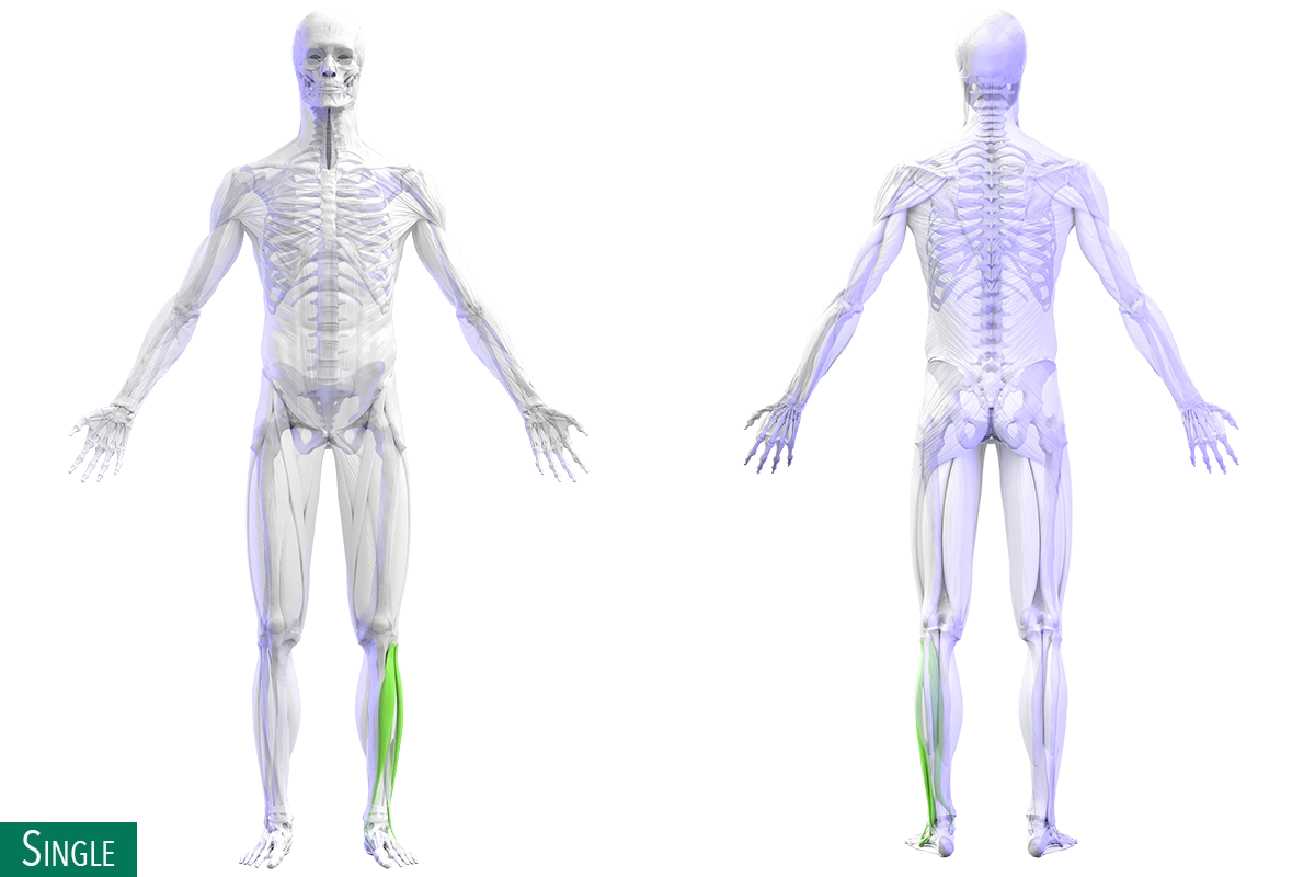

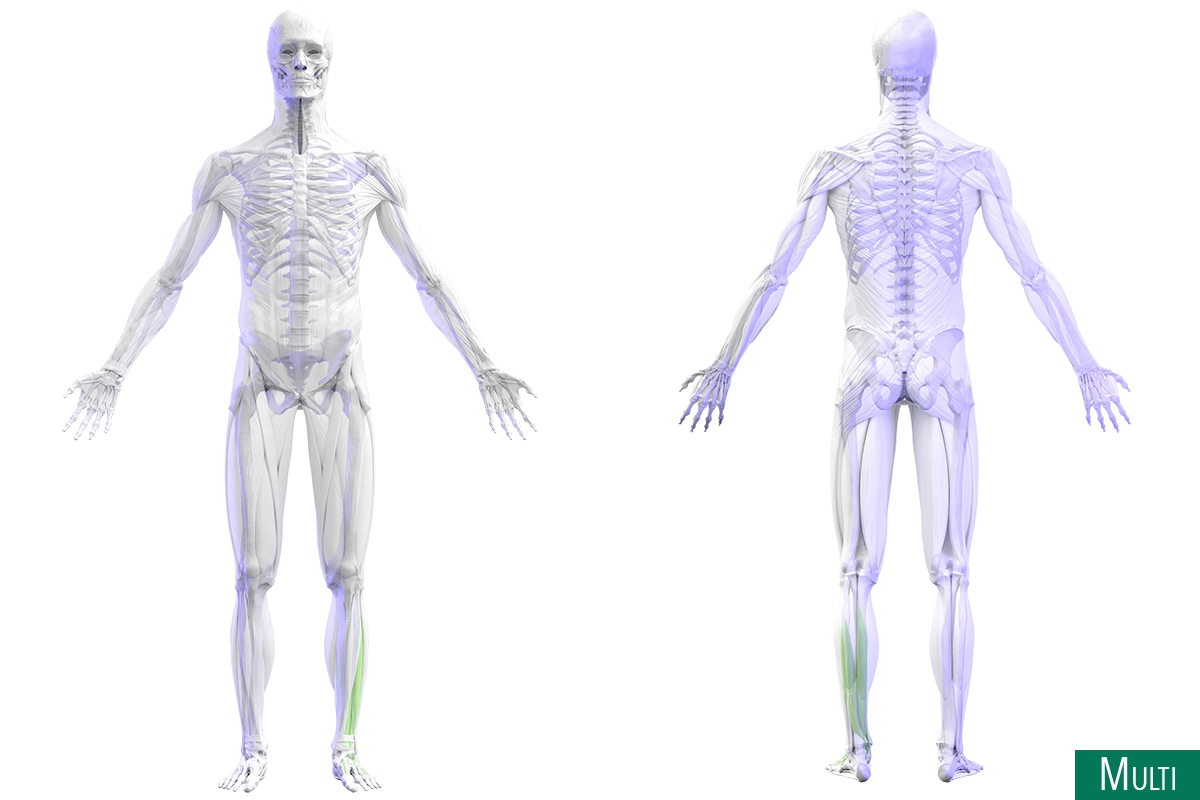
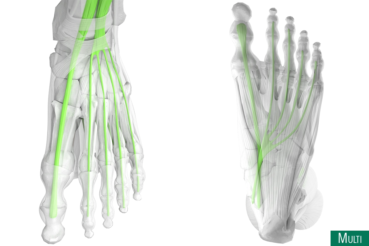

Reciprocal intra-complex relationships of the frontal ankle. Directly or indirectly, impactfull or subtle, every organ system relates to and affects the frontal ankle.
Reciprocal Intra-Complex Relationships
As mentioned previously, a complex is a cluster of directly and indirectly connected structures from all organ systems (including the fascial system) grouped by a plane-specific motion they share: flexion-extension, abduction-adduction, or lateral-medial rotation.
The structures that make up a complex (skin, joints, muscles, nerves, arteries, heart, kidneys, etc.) interconnect and maintain reciprocal relationships. In other words, a change in one structure causes a change in another within the same complex.
Reciprocal relationships within a complex transmit influence that can be positive or negative. The impact between structures may be obvious, subtle, or unnoticeable. Sometimes it is measurable (Brinjikji et al., 2015; Kannus, 1997).
Dysfunctional frontal ankle joints, for example, negatively impact the frontal ankle muscles (Rice & McNair, 2010) and skin (van Cranenburgh, 2000; Bernards, 1991). Similarly, healthy, fully functional frontal ankle muscles and skin positively affect the frontal ankle joints.
Frontal ankle:
reciprocal intra-complex relationships
* segmental link
reciprocal inter-complex relationships
Neighboring complexes interconnect because they share motion and structures across all organ systems.
Along these reciprocal inter-complex relationships, complexes exchange influence for better or worse. These relationships exist within the same plane (intra-plane) and between planes (inter-plane). Like intra-complex relationships, influences range from positive to negative. The effects can be obvious, subtle, unnoticeable, measurable, or unquantifiable (Brinjikji et al., 2015; Kannus, 1997).
Building on reciprocal inter-complex relationships, shared structures in the nervous, fascial, integumentary, skeletal, muscular, and other systems connect the sagittal big and lesser toes, the sagittal ankle, the frontal knee and hip, and the transverse knee and hip to the frontal ankle (Wainner et al., 2007; Sueki et al., 2013).
In summary, through the organ systems and the motions they generate, the sagittal big and lesser toes, the sagittal ankle, the frontal knee and hip, and the transverse knee and hip affect the frontal ankle.
Recognizing that all organ systems are involved in motion, you may focus on a few for ease and clarity. For example, emphasizing the integumentary, skeletal, and muscular systems creates a clear, practical picture. Still, since all organ systems are interconnected, those not in focus remain involved.
With all these connections in mind, how can this information help you in a clinical setting?
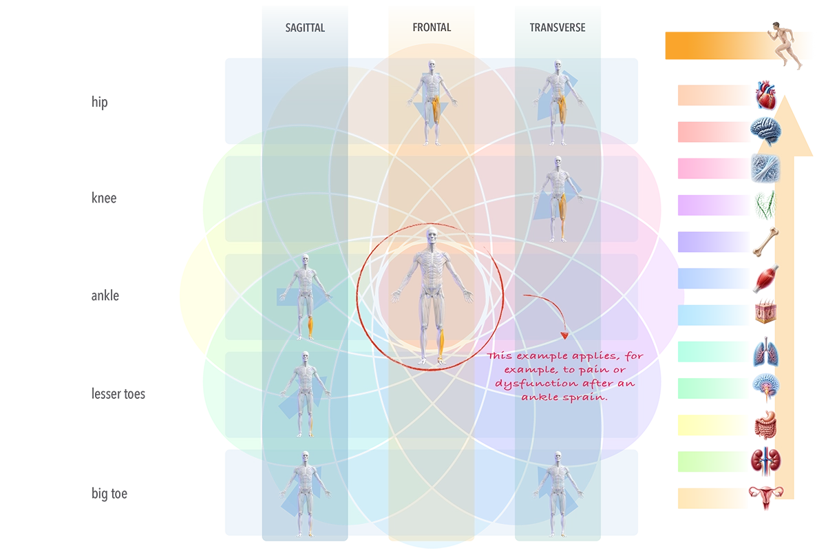
Reciprocal inter-complex relationships (50% impact and up) of the frontal ankle. Directly or indirectly, impactfull or subtle, the sagittal ankle, big and lesser toes, the transverse hip, knee, and lesser toes, and the frontal hip.
Frontal ankle:
reciprocal inter-complex relationships
Next Chapter
Summary & References
Motion Anatomy
- Any real motion (sagittal knee flexion, frontal thoracic lateral flexion, transverse shoulder rotation) involves a cluster of structures and crosses structure, region, and system.
- That's the core challenge: gross anatomy groups by structure/region/system, but motion ignores those boundaries, so to organize anatomy by motion the body must be regrouped around the structures that produce it — and that grouping is the complex.
What Is A Complex
- A complex is a cluster of interconnected structures from every organ system (plus fascia) grouped by a shared plane-specific motion — flexion-extension, abduction-adduction, or lateral-medial rotation.
- Motion happens because everything in a complex interconnects, communicates, and does its part — motion is a team effort.
- There are 19 complexes, from the big toe to the fingers; neighboring complexes may share structures, but each complex's total combination is unique.
Complexes Per Plane
- Each plane contains 19 complexes, a mix of active and passive: sagittal has 16 active and 3 passive, frontal 12 active and 7 passive, transverse 11 active and 8 passive complexes.
- Passive complexes (e.g., the passive sagittal pubic symphysis, frontal knee, transverse ankle) move only a little, and chiefly through the movement of their active neighbors.
An Active And Passive Complex
- A passive complex takes part in its plane but lacks dedicated movers and isn't built for substantial motion there; the frontal knee is the example — it moves only a few degrees in the frontal plane, restrained by its articular surfaces and the collateral and cruciate ligaments, with no abductor/adductor muscles of its own.
- It works both ways: too much frontal-plane knee motion risks ACL/MCL injury, but a passive complex that gets completely stuck also causes pain — both active and passive complexes must stay functional.
- Please note, among passive complexes, only the pubic symphysis, sacroiliac, and rib cage have well-established motion research.
Single- And Multi-Complex Muscles
- A single-complex muscle serves one complex — usually a main mover, often deep and close to the bone (vastus lateralis, gluteus medius, lumbar multifidus, brachialis).
- A multi-complex muscle serves two or more complexes — usually more superficial, helping control and coordinate (tensor fasciae latae across hip and knee; latissimus dorsi across several).
Reciprocal Intra- And Inter-Complex Relationships
- Within a complex, structures have reciprocal (give-and-take) relationships: a change in one structure changes another, and that influence ranges from positive to negative and from obvious to unnoticeable, sometimes measurable and sometimes not.
- Between complexes, shared structures create reciprocal inter-complex relationships that exchange influence both within a plane (intra-plane) and across planes (inter-plane) — for better or worse.
Reference list chapter 5
- Arndt, A., Wolf, P., Liu, A., Nester, C., Stacoff, A., Jones, R., Lundgren, P., & Lundberg, A. (2007). Intrinsic foot kinematics measured in vivo during the stance phase of slow running. Journal of Biomechanics, 40(12), 2672–2678. https://doi.org/10.1016/j.jbiomech.2006.12.009
- Athanasiou, N., Bogdanis, G. C., & Mastorakos, G. (2023). Endocrine responses of the stress system to different types of exercise. Reviews in Endocrine and Metabolic Disorders, 24(2), 251–266. https://doi.org/10.1007/s11154-022-09758-1
- Benias, P. C., Wells, R. G., Sackey-Aboagye, B., Klavan, H., Reidy, J., Buonocore, D., Miranda, M., Kornacki, S., Wayne, M., Carr-Locke, D. L., & Theise, N. D. (2018). Structure and distribution of an unrecognized interstitium in human tissues. Scientific Reports, 8, 4947. https://doi.org/10.1038/s41598-018-23062-6
- Bernards, A. T. M. (1991). Fysiologie en pathofysiologie van nocisensoriek. In H. C. F. van Zutphen, H. W. R. van Sambeek, & R. A. B. Oostendorp (Eds.), Nederlands leerboek der fysische therapie in engere zin, Deel I (4th ed.). Utrecht: Wetenschappelijke uitgeverij Bunge.
- Beyer, B., Sholukha, V., Dugailly, P. M., Rooze, M., Moiseev, F., Feipel, V., & Van Sint Jan, S. (2014). In vivo thorax 3D modelling from costovertebral joint complex kinematics. Clinical biomechanics (Bristol, Avon), 29(4), 434–438. https://doi.org/10.1016/j.clinbiomech.2014.01.007
- Brinjikji, W., Luetmer, P. H., Comstock, B., Bresnahan, B. W., Chen, L. E., Deyo, R. A., Halabi, S., Turner, J. A., Avins, A. L., James, K., Wald, J. T., Kallmes, D. F., & Jarvik, J. G. (2015). Systematic literature review of imaging features of spinal degeneration in asymptomatic populations. American Journal of Neuroradiology, 36(4), 811–816. https://doi.org/10.3174/ajnr.A4173
- Chidi-Ogbolu, N., & Baar, K. (2019). Effect of estrogen on musculoskeletal performance and injury risk. Frontiers in Physiology, 9, 1834. https://doi.org/10.3389/fphys.2018.01834
- Cranenburgh, B. van. (2000). Segmentale verschijnselen: Een bijdrage aan diagnostiek en therapie (2nd ed.). Houten: Bohn Stafleu van Loghum.
- Dempsey, J. A., La Gerche, A., & Hull, J. H. (2020). Is the healthy respiratory system built just right, overbuilt, or underbuilt to meet the demands imposed by exercise? Journal of Applied Physiology, 129(6), 1235–1256. https://doi.org/10.1152/japplphysiol.00444.2020
- Fischer, K. M., Willwacher, S., Arndt, A., & Brüggemann, G.-P. (2018). Calcaneal adduction and eversion are coupled to talus and tibial rotation. Journal of Anatomy, 233(1), 64–72. https://doi.org/10.1111/joa.12813
- Hackney, A. C., & Lane, A. R. (2015). Exercise and the regulation of endocrine hormones. Progress in Molecular Biology and Translational Science, 135, 293–311. https://doi.org/10.1016/bs.pmbts.2015.07.001
- Handelsman, D. J., Hirschberg, A. L., & Bermon, S. (2018). Circulating testosterone as the hormonal basis of sex differences in athletic performance. Endocrine Reviews, 39(5), 803–829. https://doi.org/10.1210/er.2018-00020
- Hicks J. H. (1954). The mechanics of the foot. II. The plantar aponeurosis and the arch. Journal of anatomy, 88(1), 25–30.
- Hunter, S. K., Angadi, S. S., Bhargava, A., Harper, J., Hirschberg, A. L., Levine, B. D., Moreau, K. L., Nokoff, N. J., Stachenfeld, N. S., & Bermon, S. (2023). The biological basis of sex differences in athletic performance: Consensus statement for the American College of Sports Medicine. Medicine & Science in Sports & Exercise, 55(12), 2328–2360.
- Inman, V. T. (1976). The joints of the ankle. Baltimore: Williams & Wilkins.
- Jeukendrup, A. E. (2017). Periodized nutrition for athletes. Sports Medicine, 47(Suppl 1), 51–63. https://doi.org/10.1007/s40279-017-0694-2
- Johanson, M. A., Baer, J., Hovermale, H., & Phouthavong, P. (2008). Subtalar joint position during gastrocnemius stretching and ankle dorsiflexion range of motion. Journal of Athletic Training, 43(2), 172–178.
- Kannus, P. (1997). Etiology and pathophysiology of chronic tendon disorders in sports. Scandinavian Journal of Medicine & Science in Sports, 7(2), 78–85. https://doi.org/10.1111/j.1600-0838.1997.tb00123.x
- Kraemer, W. J., & Ratamess, N. A. (2005). Hormonal responses and adaptations to resistance exercise and training. Sports Medicine, 35(4), 339–361. https://doi.org/10.2165/00007256-200535040-00004
- Lundgren, P., Nester, C., Liu, A., Arndt, A., Jones, R., Stacoff, A., Wolf, P., & Lundberg, A. (2008). Invasive in vivo measurement of rear-, mid- and forefoot motion during walking. Gait & Posture, 28(1), 93–100. https://doi.org/10.1016/j.gaitpost.2007.10.009
- McClay, I., & Manal, K. (1997). Coupling parameters in runners with normal and excessive pronation. Journal of Applied Biomechanics, 13(1), 109–124.
- Neumann, D. A. (2017). Kinesiology of the musculoskeletal system: Foundations for rehabilitation (3rd ed.). Mosby/Elsevier.
- Ormsbee, M. J., Bach, C. W., & Baur, D. A. (2014). Pre-exercise nutrition: The role of macronutrients, modified starches and supplements on metabolism and endurance performance. Nutrients, 6(5), 1782–1808. https://doi.org/10.3390/nu6051782
- Oxford English Dictionary. (n.d.). Complex (n.). In Oxford English Dictionary. Retrieved [date], from https://www.oed.com
- Poortmans, J. R., & Ouchinsky, M. (2006). Glomerular filtration rate and albumin excretion after maximal exercise in aging sedentary and active men. The Journals of Gerontology Series A: Biological Sciences and Medical Sciences, 61(11), 1181–1185. https://doi.org/10.1093/gerona/61.11.1181
- Rice, D. A., & McNair, P. J. (2010). Quadriceps arthrogenic muscle inhibition: Neural mechanisms and treatment perspectives. Seminars in Arthritis and Rheumatism, 40(3), 250–266. https://doi.org/10.1016/j.semarthrit.2009.10.001
- Romer, L. M., & Polkey, M. I. (2008). Exercise-induced respiratory muscle fatigue: Implications for performance. Journal of Applied Physiology, 104(3), 879–888. https://doi.org/10.1152/japplphysiol.01157.2007
- Sinnatamby, C. S. (2011). Last's anatomy: Regional and applied (12th ed.). Churchill Livingstone/Elsevier.
- Stacoff, A., Nigg, B. M., Reinschmidt, C., van den Bogert, A. J., Lundberg, A., Denoth, J., & Stüssi, E. (2000). Tibiocalcaneal kinematics of barefoot versus shod running. Journal of Biomechanics, 33(11), 1387–1395.
- Standring, S. (Ed.). (2021). Gray's anatomy: The anatomical basis of clinical practice (42nd ed.). Elsevier.
- Sturesson, B., Uden, A., & Vleeming, A. (2000). A radiostereometric analysis of movements of the sacroiliac joints during the standing hip flexion test. Spine, 25(3), 364–368. https://doi.org/10.1097/00007632-200002010-00018
- Sueki, D. G., Cleland, J. A., & Wainner, R. S. (2013). A regional interdependence model of musculoskeletal dysfunction: Research, mechanisms, and clinical implications. Journal of Manual & Manipulative Therapy, 21(2), 90–102. https://doi.org/10.1179/2042618612Y.0000000027
- Wainner, R. S., Whitman, J. M., Cleland, J. A., & Flynn, T. W. (2007). Regional interdependence: a musculoskeletal examination model whose time has come. The Journal of orthopaedic and sports physical therapy, 37(11), 658–660. https://doi.org/10.2519/jospt.2007.0110
- Walheim, G. G., & Selvik, G. (1984). Mobility of the pubic symphysis: In vivo measurements with an electromechanic method and a roentgen stereophotogrammetric method. Clinical Orthopaedics and Related Research, 191, 129–135.
- Young, R., Nix, S., Wholohan, A., Bradhurst, R., & Reed, L. (2013). Interventions for increasing ankle joint dorsiflexion: a systematic review and meta-analysis. Journal of Foot and Ankle Research, 6(1), 46. https://doi.org/10.1186/1757-1146-6-46
Move toward a better you!
Strength
Flexibility

Biological Age
A Stronger Heart

Mental Resilience

Sharper Mind

Stronger Bones